
Checking Your Horses Health with Ultrasound Imaging
There are instances where radiographs alone cannot diagnose a problem or fully reveal the issues contributing to a single lameness or disease. Soft tissue injuries are common and require more specialized diagnostic imaging for which ultrasound is the mainstay in equine ambulatory medicine.
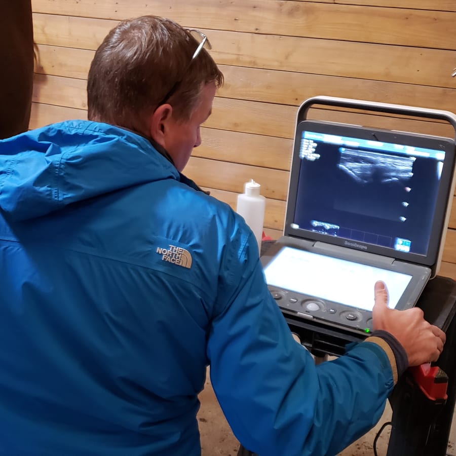
uses high-resolution portable ultrasound machines, giving us the capability to evaluate tendons, ligaments, and joint structures in the field. For emergencies, the ultrasound is invaluable in the assessment of colics and other illnesses such as pneumonia and peritonitis too.
Below are several instances of its use in lameness, however, this machine can help us diagnose causes of colic, pneumonia, abscesses, and the list goes on. Having this technology brings another level of diagnostics on par with what clients receive at referral practices.
Ultrasound Case Studies
We are committed to educating our clients about their horse's health. If you are interested in learning more about what our veterinarians can see with ultrasounds, take a look at the case studies below.
Normal Medial Meniscus
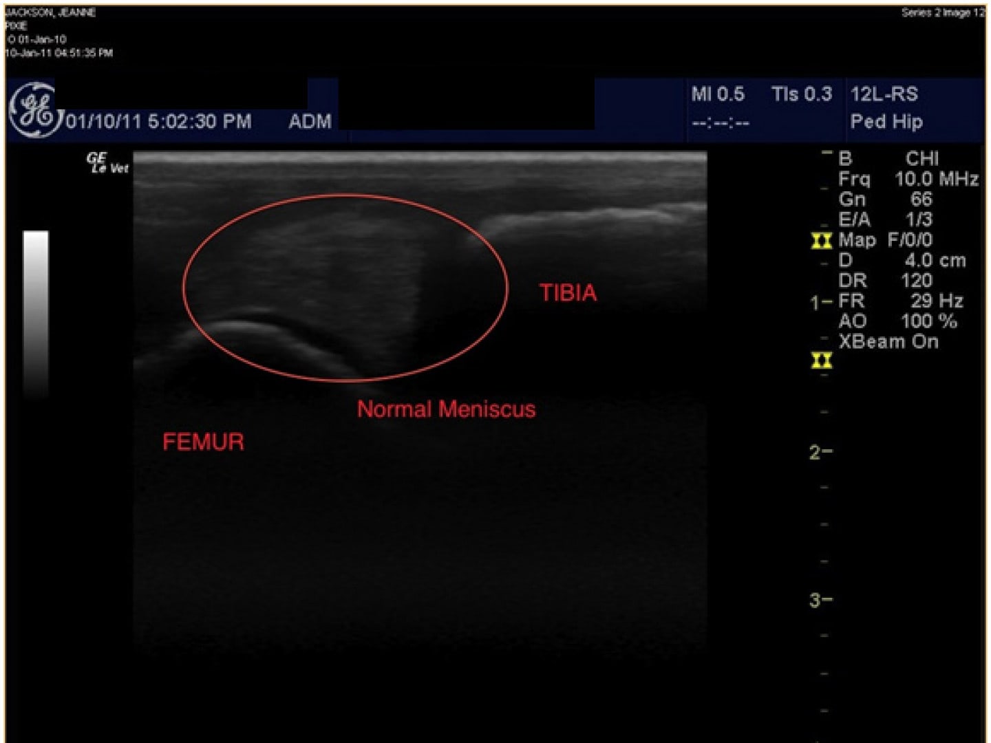
This is an ultrasonic image of a normal medial meniscus in the stifle of a normal, sound horse. The meniscus has normal consistency throughout the tissue with no visible tears or disruptions.
Meniscus With Hind Limb Lameness
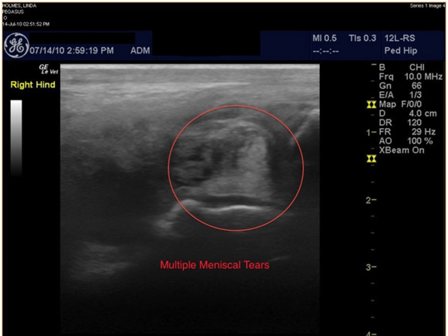
Using ultrasound, we were able to diagnose multiple meniscal tears that were not visible on X-rays. These tears caused significant instability and pain in that joint. The prognosis for this type of injury is typically poor.
Cartilage
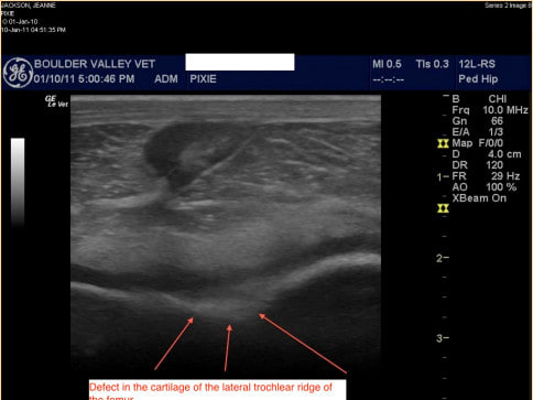
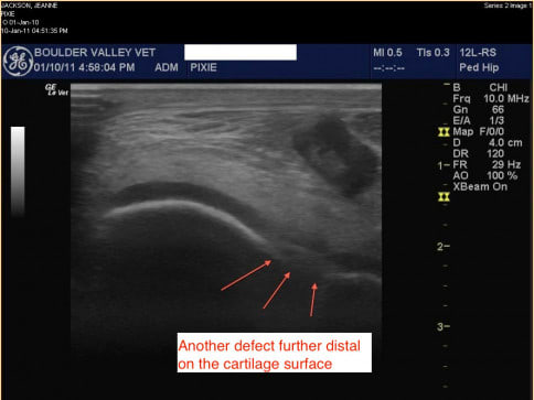
These two images show the cartilage along the trochlear ridges of the femur in the stifle of a 6-month old foal with acute lameness and effusion in the joint. X-rays of this joint could not demonstrate any changes in the cartilage of the joint. The ultrasound machine was used to visualize the cartilage, which is represented by the thick dark line along the bone which is the bright white line. The arrows show areas where there are cartilage defects. These are OCD lesions and the horse was sent to surgery. In surgery, multiple cartilage flaps and erosion's were visualized and cleaned up.